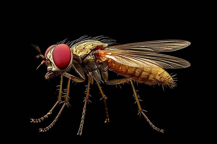
Introduction: Overview of Drosophila Brain Research
Drosophila melanogaster, commonly known as the fruit fly, has long been a cornerstone of genetic research, but its implications in understanding neural circuits are gaining unprecedented attention. With an intricately organized brain that boasts about 100,000 neurons, Drosophila provides a simplified yet powerful model for unraveling complex brain functions and behaviors. Recent advances in neuroimaging techniques and genome editing have allowed researchers to visualize neural activity with remarkable precision, revealing not just structures but also dynamic networks that underpin sensation, decision-making, and memory.
Moreover, the exploration of Drosophila’s connectome—an exhaustive map of neural connections—offers fresh insights into how specific neuron types interact within the circuit. This endeavor not only enhances our understanding of basic biological processes but also serves as a crucial comparison point for vertebrate neurological studies. As scientists delve deeper into this miniature brain’s architecture and functioning through innovative labeling techniques and cell classification systems, they pave the way toward deciphering human neurological disorders by highlighting conservation across species. In essence, Drosophila stands at the intersection of genetics and neuroscience—a vital contributor to our quest for knowledge about cognition and behavior on both fundamental biological levels and potential clinical applications.
Importance of Brain Labeling Techniques
Brain labeling techniques are crucial for advancing our understanding of neural structures and functions, especially in model organisms like Drosophila. These innovative methodologies allow researchers to map out the intricate architecture of the brain, facilitating insights into its diverse connectivity patterns. By employing techniques such as transgenic labeling or viral tracing, scientists can visualize specific neuron types and their synaptic interactions in unprecedented detail.
The significance of these techniques extends beyond mere anatomical mapping; they offer a gateway to unraveling the complexities of neural circuits and behaviors. For instance, by pinpointing active pathways during certain behavioral tasks, we gain valuable perspectives on how these circuits interact to generate specific outcomes. Furthermore, as we explore variations in connectomes across different genetic backgrounds or environmental contexts, brain labeling empowers researchers to make critical correlations between structure and function—a key step towards deciphering not only Drosophila’s biology but also broader principles that may apply across species.
Methods for Cell Classification in Drosophila
Cell classification in Drosophila has evolved dramatically with the advent of advanced imaging techniques and machine learning algorithms. Researchers are now leveraging high-resolution microscopy combined with computational approaches to pinpoint cellular identities within complex brain structures. For instance, methods such as single-cell transcriptomics not only provide a detailed profile of gene expression but also enable the identification of previously unrecognized cell types based on subtle variations in their molecular signatures. This could lead to newfound insights into the roles these cells play in neural circuitry and behavior.
Moreover, clustering techniques, like t-distributed stochastic neighbor embedding (t-SNE) and Uniform Manifold Approximation and Projection (UMAP), empower scientists to visualize vast datasets in intuitive ways by grouping similar cell populations based on multi-dimensional features. Coupled with deep learning frameworks trained on annotated images, researchers can achieve unprecedented accuracy in classifying cells under varying conditions or stressors, revealing dynamic responses that were previously hidden from view. This synergy between biological intuition and computational power paves the way for groundbreaking discoveries about Drosophila’s neural connectivity and its implications for understanding more complex organisms.
The Role of Connectomics in Neuroscience
Connectomics, the mapping of neural connections within the brain, is revolutionizing our understanding of neuronal architecture and function. By meticulously tracing the intricate web of synaptic connections, researchers can uncover not only how information is processed but also how behavioral patterns emerge from complex neural interactions. In species like Drosophila, where a more straightforward nervous system allows for high-resolution mapping, scientists are poised to decipher the fundamental principles governing connectomic structures that could be analogous across more complex organisms.
Importantly, connectomics bridges various disciplines within neuroscience—from genetics and molecular biology to computational modeling—enabling a holistic view of brain networks. Recent advancements in high-throughput imaging techniques have accelerated the identification and classification of diverse cell types based on their connectivity patterns. This has profound implications; understanding these relationships sheds light on developmental processes and potentially unveils targets for therapeutic intervention in neurodegenerative diseases or psychiatric disorders. As we untangle these connections, we begin to see the symphony of neuronal communication that underlies cognition and behavior—a truly captivating window into the biological intricacies that define sentient life.
Key Findings from Recent Studies
Recent studies have illuminated the intricate tapestry of neural connections in Drosophila, revealing a labyrinth of cell types and connectivity patterns that challenge our previous understanding. One significant finding is the identification of novel neuron classes that play unique roles in processing sensory information. By employing cutting-edge imaging techniques coupled with advanced machine learning algorithms, researchers were able to map these neurons’ distinct firing patterns in response to various stimuli, underscoring the complexity of behavioral responses orchestrated by simple circuit structures.
Moreover, connectome analyses have unveiled surprising similarities between Drosophila and higher organisms, including humans. This raises intriguing questions about evolutionary conservation and functional parallels across species. The studies highlight how even minor structural variances can lead to substantial differences in behavior, suggesting that the evolutionary pressures on neural circuitry may be sharper than previously thought. Such insights not only enhance our foundational understanding of brain architecture but also pave the way for future research aimed at uncovering the molecular underpinnings driving these behaviors, offering tantalizing prospects for advancements in neurobiology and beyond.
Challenges in Brain Mapping and Classification
The intricacies of brain mapping and the classification of neural cell types pose significant challenges that extend beyond mere technical hurdles. One major issue is the sheer complexity of neuronal connectivity within the Drosophila brain, where myriad synaptic connections intricately intertwine. This tangled web can obscure our understanding, making it difficult to delineate functional circuits accurately. Moreover, as we grapple with advanced imaging techniques and big data analytics, the risk of data interpretation bias becomes a pressing concern, necessitating refined algorithms that account for variability in neuron morphology and behavior.
Moreover, current classification frameworks often rely on traditional histological methods or simplistic morphological criteria, which may fail to capture the dynamic and context-dependent nature of neuron function. As we move toward a more integrated approach that combines genomic information with functional data, new classification systems will need to evolve rapidly. It’s essential that these systems embrace neuroethology—understanding neurons in their ecological context—to fully appreciate how diverse cell types interact in real-time behaviors. This paradigm shift not only enhances our comprehension of Drosophila but also lays foundational knowledge applicable to more complex organisms, thereby broadening the implications for neuroscience as a whole.
Tools and Technologies Used in Research
In the ever-evolving landscape of neuroscience, the tools and technologies employed in research are crucial for unraveling the complexities of brain structure and function. Advanced imaging techniques, such as two-photon microscopy and serial block-face electron microscopy, allow researchers to visualize neuronal circuits in exquisite detail. These methods not only enable high-resolution imaging but also facilitate dynamic observation of neural activity over time, revealing insights into how connections strengthen or weaken with experience. The integration of these powerful imaging modalities with genetic tools like CRISPR-Cas9 allows for precise manipulation of specific cell types within the Drosophila brain, thus providing unprecedented opportunities for analyzing connectomic landscapes.
Moreover, machine learning algorithms have become indispensable in processing vast datasets generated by high-throughput sequencing and imaging technologies. These smart computational approaches can automate the classification of diverse neural cell types based on their morphological features and connectivity patterns, significantly accelerating data analysis and interpretation. By leveraging artificial intelligence to identify subtle differences between neuronal populations, researchers can uncover novel relationships between form and function that were previously inaccessible. This intersection of biology with cutting-edge technology is not just propelling our understanding forward; it is redefining what we believe possible in neuroscience research.
Future Directions for Drosophila Connectome Studies
As researchers unlock the intricacies of Drosophila connectomes, future directions seem poised to embrace advancements in artificial intelligence and machine learning. These technologies can enhance data analysis, allowing scientists to decipher complex neural patterns with unprecedented precision. Imagine algorithms that not only identify structural connections within the brain but also predict functional outcomes based on these relationships. This integration could revolutionize our understanding of how neural circuits govern behavior.
Moreover, the emerging field of optogenetics combined with advanced imaging techniques promises to take connectome studies a step further. By precisely manipulating neuron activity while capturing real-time responses in live fly models, researchers will be able to establish causal links between specific synaptic interactions and behavioral phenomena. As we venture into personalized neurogenetics, tailoring studies based on individual genetic backgrounds could yield insights into variability in neural processing among different flies, paving the way for nuanced investigations into neurodevelopmental disorders and cognitive processes across species.
Implications for Understanding Human Brain Disorders
The development of a comprehensive labeling system for the Drosophila brain, intertwined with advanced connectome cell classification, offers profound implications for understanding human brain disorders. By leveraging the simplicity of the fruit fly’s neural architecture, researchers can unravel the complex wiring and functional networks that may be analogous to those in humans. This approach not only streamlines experimental processes but also enables the identification of conserved pathways crucial in neurodevelopmental and neurodegenerative diseases.
Furthermore, as we parse out specific neural circuits linked to behaviors in Drosophila, there lies immense potential to pinpoint corresponding mechanisms in human pathologies. For instance, insights gained from studying synaptic connections underlying memory or anxiety responses can illuminate similar frameworks disrupted in conditions like Alzheimer’s or depression. Ultimately, this research paradigm fosters a richer dialogue between model organisms and human health—a bridge that enhances our comprehension of intricate psychiatric disorders and guides therapeutic innovations tailored to restore normal brain function.
Conclusion: Significance of Comprehensive Research Insights
The significance of comprehensive research insights in the study of Drosophila brain labeling and connectome cell classification cannot be overstated. By meticulously mapping neural circuits and identifying distinct cell types, researchers unveil the complex tapestry of neurobiological function that governs behavior and cognition. This multifaceted approach not only facilitates a deeper understanding of Drosophila’s neural architecture but also serves as a proxy for unraveling similar mechanisms in more complex organisms, including humans.
Moreover, these insights pave the way for innovative methodologies in neuroscience. The integration of advanced imaging techniques with machine learning algorithms promises unprecedented accuracy in data analysis and interpretation. As we glean more about how various neuronal populations interact within the connectome framework, there is potential for breakthroughs in neurodevelopmental disorders and regenerative medicine. Ultimately, such comprehensive research efforts not only illuminate fundamental principles of brain organization but also inspire new avenues for therapeutic interventions across species boundaries.

